Warning Signs of Eye Trouble
 |
Color Blindness Test
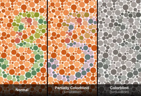 |
Which number do you see in the far left? If it's "3," you probably have normal color vision. If it's a "5," you may be colorblind. This view is simulated in the center panel and represents a mild color vision deficiency. About 10% of men are born colorblind, but few women. Complete color blindness (very rare) is simulated at right. No number is visible. Tinted glasses may help the colorblind see better.
Nearsightedness (Myopia)
About 33% of Americans (ages 12-54) have a blurry view of distant objects, called myopia, up from about 25% in the early 1970s. Risk factors include:
- Family history (one or both parents)
- Lots of prolonged, close-up reading
Farsightedness (Hyperopia)
Most of us are born with mild farsightedness, but normal growth in childhood often corrects the problem. When it persists, you may see distant objects well, but books, knitting, and other close objects are a blur. Hyperopia runs in families. Symptoms include trouble with reading, blurry vision at night, eyestrain, and headaches. It can be treated with glasses, contacts, or surgery in some cases.
Presbyopia
Just like gray hair or wrinkles, trouble reading fine print is a sign of aging. Called presbyopia -- or "old eye" in Greek -- symptoms appear in the 40s. The eyes' lenses become less flexible and can't change shape to focus on objects at reading distance. The solution: reading glasses or bifocals, which correct both near and distance vision. If you wear contacts, ask your eye doctor about contacts made for people with presbyopia.
Nearsightedness: What Happens
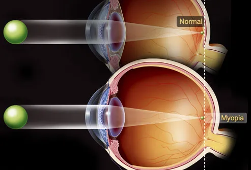
Typically, an eyeball that's too long causes myopia. But an abnormally shaped cornea or lens can also be to blame. Light rays focus just in front of the retina, instead of directly on it. This sensitive membrane lines the back of the eye (seen in yellow) and sends signals to the brain through the optic nerve. Nearsightedness often develops in school-age children and teens, who need to change glasses or contacts frequently as they grow. It usually stabilizes by the early 20s.
Read Also: Exercising Safely With Allergic Asthma
Farsightedness: What Happens
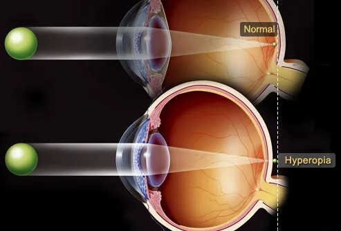
In hyperopia, the cause is often an eyeball that is too short. Light rays focus behind the retina, causing close objects to be blurry. In severe cases of hyperopia, especially after the age of 40, distance vision can be blurred as well. An abnormal shape in the cornea or lens can also lead to farsightedness. Children with significant hyperopia are more likely to have crossed eyes (strabismus) or lazy eye (amblyopia) and may have difficulty reading. That’s one of the reasons eye doctors recommend vision exams for young children.
Astigmatism
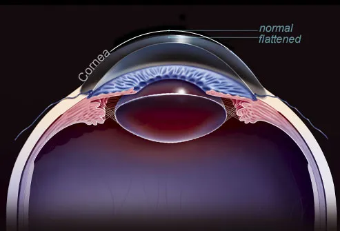 |
Your vision may be out of focus at any distance with an astigmatism in one or both eyes. It occurs when the cornea, the clear “window” that covers the front of the eye, is misshapen. Light rays can be scattered in different points on the retina, rather than focusing on a single point. Glasses or contact lenses correct the problem, and surgery may be another option. Along with blurred vision, symptoms may include headaches, fatigue, and eye strain.
Refractive Eye Surgery
Do you dream of seeing clearly without glasses? Surgery to reshape the cornea can correct nearsightedness, farsightedness, or astigmatism with a success rate of better than 90%. People with severe dry eye, thin or abnormally shaped corneas, or severe vision problems may not be good candidates. Possible side effects include glare or sensitivity to light.
Glaucoma: View
 |
You can't feel it, but deterioration of the optic nerve oftentimes with elevated eye pressure can silently steal your sight, a condition called glaucoma. There may be no symptoms until central vision is lost (following gradual loss of peripheral vision), so regular eye exams are critical to find it early. Those at higher risk include:
- African-Americans over 40
- Anyone over 60, especially Mexican-Americans
- People with a family history
Glaucoma: What Happens
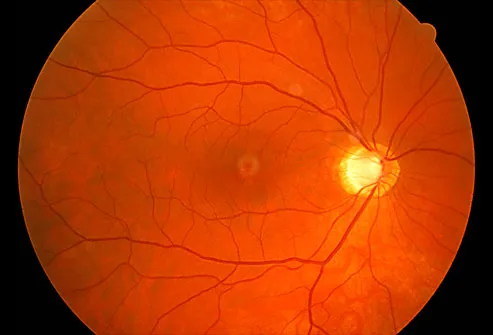 |
In the most common form of glaucoma, increased eye pressure leads to optic nerve damage and loss of vision. The eye is filled with circulating fluid that nourishes its internal structures. Sometimes the balance between fluid creation and exit is abnormal. The buildup of fluid increases pressure and damages the optic nerve at the back -- the bundle of 1 million nerve fibers that carry information to the brain. Without treatment, glaucoma can cause total blindness.
The bright yellow circle shows an optic nerve head that is damaged by glaucoma. The dark central area is the macula, responsible for finely-detailed central vision.
Read Also: Spring Allergies
Macular Degeneration: View

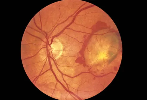
Age-related macular degeneration (AMD) damages, then destroys, the eye's finely-detailed central vision, making it difficult to read or drive. Symptoms can include a central blurry spot or straight lines that appear wavy. Finding and treating AMD promptly can help slow vision loss. Being over 60, smoking, high blood pressure, obesity, and a family history of AMD increase your risk.
Macular Degeneration: What Happens
 |
Macular Degeneration: Test

Cover one eye and stare at the center dot in this Amsler Grid, from a distance of 12 to 15 inches. (You can wear your reading glasses.) Do you see wavy, broken, or blurry lines? Are any areas distorted or missing? Repeat the procedure for your other eye. While no self-test can substitute for an eye exam, this grid is used to help detect early symptoms of AMD
Macular Degeneration:Signs
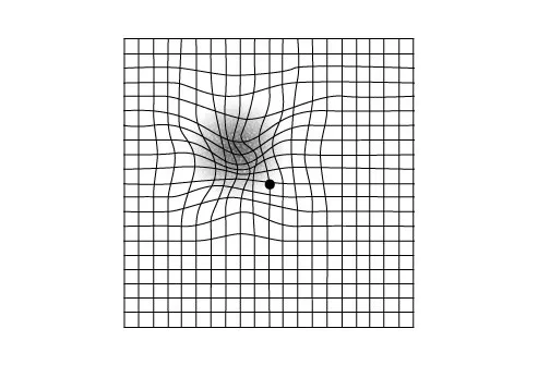 |
As seen here, the Amsler Grid can look quite distorted to someone with significant macular degeneration and may include a central dark spot. Straight lines that appear wavy are also cause for concern, as they can be an early symptom of "wet" AMD, the more serious and fast-moving type of macular degeneration. Your eye care professional will want to evaluate you right away, starting with a thorough dilated eye exam.
 |
| \ |
Diabetic Retinopathy: View
Type 1 and type 2 diabetes can cause partial vision loss (seen here) and lead to blindness. The damage involves tiny blood vessels in the retina and can often be treated, but don't wait for symptoms. By the time they occur -- blurry vision, spots, shadows, or pain -- the disease may be severe. People with known diabetes need annual eye exams, sometimes even more often if diabetic eye changes have begun. The best prevention is keeping your blood sugar in check.
Diabetic Retinopathy: What Happens
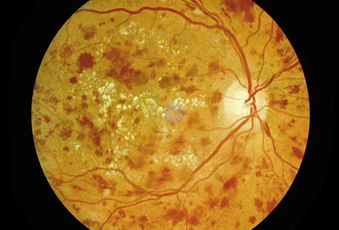 |
When high blood sugar levels go unchecked, it can damage the tiny blood vessels that support the retina. These blood vessels can swell, break, and leak fluid. In some cases, dozens of new, abnormal blood vessels grow, a condition called proliferative retinopathy. The abnormal vessels are very fragile and break open easily. These processes gradually damage the retina, causing blurred vision, blind spots, or blindness.
Cataracts: View
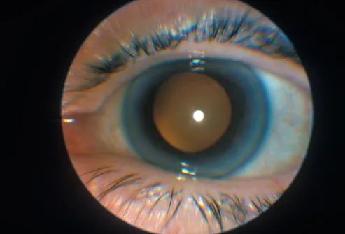
Age is not kind to our eyes. By the time we're 80 years old, more than half of us will have had a cataract, or clouding of the lens. Vision gradually gets foggy and makes it hard to read, drive, and see at night. Diabetes, smoking, or prolonged sunlight exposure may increase the risk. Surgery that replaces the clouded lens with an artificial lens is highly effective.
Read Also: Spring Allergies
Cataracts: What Happens
 |
Retinitis Pigmentosa
 |
RP is an inherited disorder that often begins with night vision problems, followed by a gradual loss of side vision, developing into tunnel vision, and finally, in some cases, blindness. One in 4,000 American have RP. A promising study showed that high-dose vitamin A supplements can reduce vision loss. However, you should consult a health care expert before taking supplements because too much vitamin A can be toxic.
Retinitis Pigmentosa: What Happens
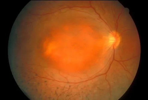 |
The light-sensitive tissue of the retina slowly deteriorates over many years in people with RP. As this tissue dies, it stops sending signals to the brain, and some vision is lost. Eye exams show abnormal dark spots (pigments) sprinkled around the retina. Early cataracts can also occur, as well as a swelling of the retina called macular edema (the central orange mass seen here).
Floaters and Specks
Blurry spots or specks in your vision that move may be floaters -- debris in the eye's vitreous gel. They don't block vision and are more easily seen in bright light. Floaters are common and usually harmless. But if they appear or increase suddenly, or are accompanied by light flashes, you should see a doctor. Vision abnormalities that tend to be more serious include persistent white or black spots and a sudden shadow or loss of peripheral vision. These require immediate evaluation.
Amblyopia (Lazy Eye)
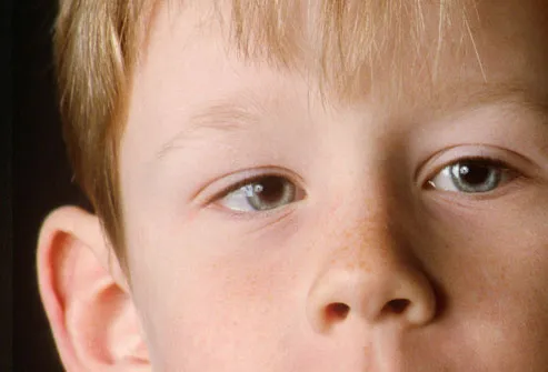 |
During childhood, when vision is reduced in one eye, the brain sometimes favors the other eye. This condition, called amblyopia, may stem from a misalignment of the eyes (strabismus or crossed eyes) or poorer vision in one eye. A patch or drops that blur the vision in the "good" eye can prod the brain to use the other eye. If untreated during childhood, vision loss from amblyopia can be permanent.
Eye Care: Object in the Eye
Many nerve endings lurk just beneath the surface or your cornea, so a tiny speck can be surprisingly painful. Don't rub the eye, or you may cause serious damage. Gently flushing the eye with lukewarm water is often recommended. If it doesn't dislodge the foreign body, or you have any questions, call a medical provider who can remove the object and provide antibiotic drops to protect the cornea from infection.
Eye Care: Tears and Dry Eye
Tears are the lubrication for our eyes. When not enough flow, perhaps due to dry air, aging, or other health conditions, the eyes can become painful and irritated. For people with mild cases of dry eye, occasionally using eye drops labeled artificial tears may do the trick. Others with more pronounced and frequent dry eye symptoms may need other medications or a procedure to block the exit ducts.
Eye Care: Pinkeye
Pinkeye, or conjunctivitis, is an inflammation caused by a virus, bacteria, irritant, or allergy. Along with the telltale redness, you might have an itching or burning sensation and a discharge. If itching is the main symptom, the cause is likely to be allergy. Most cases of infectious pinkeye are viral, which don't require antibiotics. Bacterial conjunctivitis is typically treated with antibiotic eye drops. Bacterial and viral conjunctivitis are very contagious, so wash your hands frequently while you wait for it to clear up.
Eye Care: Stye
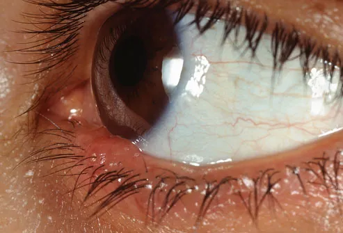 |
A stye is a tender red bump that looks like a pimple on or near the edge of the eyelid. It is just one type of infection of the eyelids (blepharitis). Styes usually heal in a week, but using a very warm, wet compress three to six times a day can speed the healing. Don’t wear contact lenses or eye makeup until it heals.
Eye Care: Allergies
Millions of Americans suffer from allergies -- and watery, itchy eyes. Pollen, grass, dust, weeds, and pet dander are common triggers. Reduce the allergens in your home with allergen impermeable covers on mattresses and pillows, thorough cleaning, and allergen filters in the furnace and air conditioner. Allergy eye drops, artificial tears, and antihistamines also may soothe the symptoms.
Eye Care: Regular Exams
Everyone needs regular eye exams, starting before the school years. This is particularly important if you have risk factors or a family history of eye problems. Beyond vision issues, the eyes can reveal underlying health problems, such as diabetes and high blood pressure, or serious disorders like stroke or brain tumor. Bulging eyes are a sign of thyroid disease, and a yellow tint of the whites of the eyes may indicate liver problems.
Eye Protection: Sun
UV rays can damage your eyes, just as they do your skin. Regular overexposure to sun can cause cataracts 8-10 years early, and a single lengthy exposure can actually burn your corneas. The solution is sunglasses that block UV rays and a hat. People with light-colored eyes are likely to have a greater sensitivity to light. New or pronounced sensitivity to bright light can be a sign of a more serious eye condition.
Eye Protection: Everyday Hazards
Grease splatters from a pan, yard debris flies up from the lawn mower, cleaning solution splashes in a bucket. Some of the greatest hazards to the eyes are in the home. Eye care specialists recommend that every household have ANSI-approved protective eyewear. Even if an eye injury seems minor, go to the emergency room immediately to check it out.
Foods for Eye Health
 |
Carrots really are good for your eyes. So are spinach, nuts, oranges, beef, fish, whole grains, and many other foods in a healthy diet. Look for foods with antioxidants such as omega-3 fatty acids; vitamins C, E, and beta-carotene; as well as zinc, lutein, and zeaxanthin. Research suggests those nutrients may reduce the risk of age-related macular degeneration.
Read Also:

No comments:
Post a Comment